
Nishant Sinha, PhD
Dr. Nishant Sinha, Assistant Professor of Informatics in the Department of Biostatistics, Epidemiology, and Informatics, is a neuroscientist with interdisciplinary expertise in engineering, informatics, biostatistics, and neurology, specializing in translational […]

Zhi Huang, PhD
Dr. Huang is an Assistant Professor of Pathology and Laboratory Medicine, with a secondary appointment in the Department of Biostatistics, Epidemiology, and Informatics at the University of Pennsylvania Perelman School of Medicine. […]
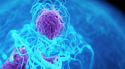
AI Tool Analyzes 30K Data Points Per Medical Imaging Pixel in Cancer Search
Mingyao Li, PhD and her Penn Medicine colleagues developed an AI-powered tool called MISO (Multi-modal Spatial Omics) that can detect cell-level characteristics of cancer by looking at data from extremely small pieces of tissue—some as small as the width of five human hairs.

Jarcy Zee, PhD
Dr. Zee’s research focuses on developing and applying methods for observational and survival data analysis and identifying novel biomarkers of kidney disease progression. She collaborates on clinical research projects in […]

Russell T. Shinohara, PhD
Dr. Shinohara’s methodological research centers on the assessment of structural and functional changes in the brain throughout the lifespan and in neurological, psychiatric, and developmental disorders. He is interested in […]

Haochang Shou, PhD
Dr. Shou’s methodological research focuses mainly on developing novel functional data analysis and machine learning models for multimodal and longitudinal imaging measures, wearable computing sensor data, and assessing biomarker reproducibility. […]

Kristin A. Linn, PhD
Dr. Linn’s methodological research addresses barriers to incorporating complex, high-dimensional data into models for personalized medicine and biomarker development using neuroimaging data. As a co-investigator, she provides biostatistical leadership on […]

Jeffrey S. Morris, PhD
In addition to his professorial role, Dr. Morris serves as Director of the Division of Biostatistics. His research interests focus on developing quantitative methods to extract knowledge from biomedical big […]

Wensheng Guo, PhD
Dr. Guo joined the Biostatistics faculty at the University of Pennsylvania in 1998, and was promoted to Professor of Biostatistics in 2010. He was elected as ASA fellow in 2010 […]

Quy Cao, PhD
Dr. Cao has established himself as a leader in the field of collaborative analytics, particularly in high-dimensional and imaging science. He has worked on problems in the study of the […]
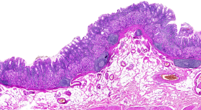
Dr. Mingyao Li Explores the Transformative Role of AI in Spatial Omics Research in Nature Methods
Dr. Mingyao Li's article in Nature Methods discusses how artificial intelligence is revolutionizing spatial omics, enhancing integration of diverse data and accelerating biological discoveries for improved health outcomes in biomedical research.

Russell Shinohara Selected for the 2023 Mortimer Spiegelman Award
Russell “Taki” Shinohara, PhD was selected as the 2023 Mortimer Spiegelman Award recipient by the Applied Public Health Statistics Section of the American Public Health Association (APHA). Each year, the APHA […]
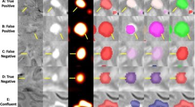
Fully Automated Detection of Paramagnetic Rims in Multiple Sclerosis Lesions on 3T Susceptibility-Based MR Imaging
The presence of a paramagnetic rim around a white matter lesion has recently been shown to be a hallmark of a particular pathological type of multiple sclerosis lesion. Increased prevalence […]
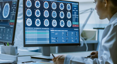
Removal of Scanner Effects in Covariance Improves Multivariate Pattern Analysis in Neuroimaging Data
To acquire larger samples for answering complex questions in neuroscience, researchers have increasingly turned to multi-site neuroimaging studies. However, these studies are hindered by differences in images acquired across multiple […]
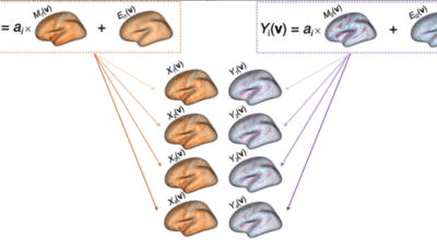
A simple permutation-based test of intermodal correspondence
While many findings in neuroimaging studies pertain to multiple imaging modalities, statistical methods underlying intermodal comparisons have varied. In our recently published paper in Human Brain Mapping, we propose the […]
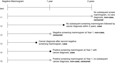
Breast Cancer Diagnosis in the ‘Between’ Year: Body Mass Index Matters
Although mammography reduces breast cancer mortality by 15 to 20 percent, the diagnosis in many cases — approximately 15 percent of all breast cancers — occurs after a patient has […]
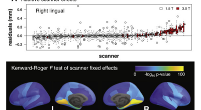
Longitudinal ComBat: A method for harmonizing longitudinal multi-scanner imaging data
While aggregation of neuroimaging datasets from multiple sites and scanners can yield increased statistical power, it also presents challenges due to systematic scanner effects. This unwanted technical variability can introduce […]
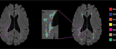
Automatic Threshold Detection for Classification of Lesions
Total brain white matter lesion volume is the most widely established MRI outcome measure in studies of multiple sclerosis. To estimate white matter lesion volume, there are a number of […]
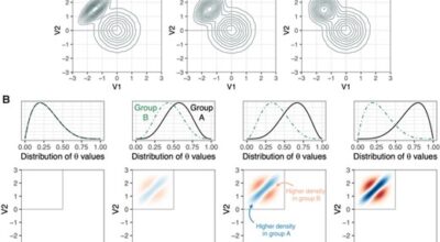
A local group differences test for subject-level multivariate density neuroimaging outcomes
A great deal of neuroimaging research focuses on voxel-wise analysis or segmentation of damaged tissue, yet many diseases are characterized by diffuse or non-regional neuropathology. In simple cases, these processes […]
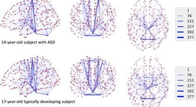
Distance-Based Analysis of Variance for Brain Connectivity
The field of neuroimaging dedicated to mapping connections in the brain is increasingly being recognized as key for understanding neurodevelopment and pathology. Networks of these connections are quantitatively represented using […]
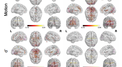
Robust Spatial Extent Inference with a Semiparametric Bootstrap Joint Inference Procedure
Spatial extent inference (SEI) is widely used across neuroimaging modalities to adjust for multiple comparisons when studying brain-phenotype associations that inform our understanding of disease. Recent studies have shown that […]
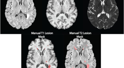
Detecting Black Holes in Multiple Sclerosis
Magnetic resonance imaging (MRI) is crucial for in vivo detection and characterization of white matter lesions (WML) in multiple sclerosis (MS). The most widely established MRI outcome measure is the […]
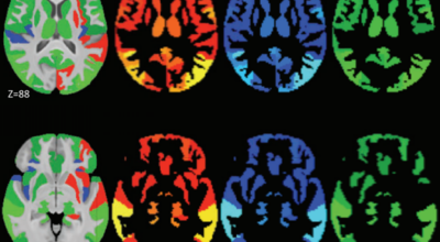
Faster family-wise error control for neuroimaging with a parametric bootstrap
In neuroimaging, hundreds to hundreds of thousands of tests are performed across a set of brain regions or all locations in an image. Recent studies have shown that the most […]
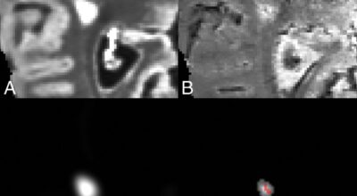
Automated Integration of Multimodal MRI for the Probabilistic Detection of the Central Vein Sign in White Matter Lesions
The central vein sign is a promising MR imaging diagnostic biomarker for multiple sclerosis. Recent studies have demonstrated that patients with MS have higher proportions of white matter lesions with […]
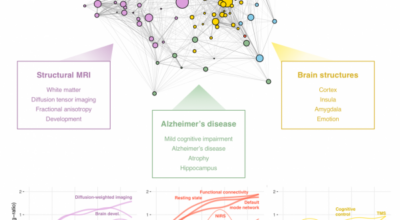
The landscape of NeuroImage-ing research
As the field of neuroimaging grows, it can be difficult for scientists within the field to gain and maintain a detailed understanding of its ever-changing landscape. While collaboration and citation […]
Interpretable High-Dimensional Inference Via Score Projection With an Application in Neuroimaging
In the fields of neuroimaging and genetics, a key goal is testing the association of a single outcome with a very high-dimensional imaging or genetic variable. Often, summary measures of […]

Detecting Multiple Sclerosis Lesions via Intermodal Coupling Analysis
Magnetic resonance imaging (MRI) is crucial for in vivo detection and characterization of white matter lesions (WML) in multiple sclerosis. While WML have been studied for over two decades using […]
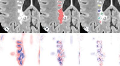
An Automated Statistical Technique for Counting Distinct Multiple Sclerosis Lesions
This study introduces a novel technique for counting pathologically distinct lesions using cross-sectional data, and demonstrates its ability to recover obscured longitudinal information. The proposed count works by incorporating information […]
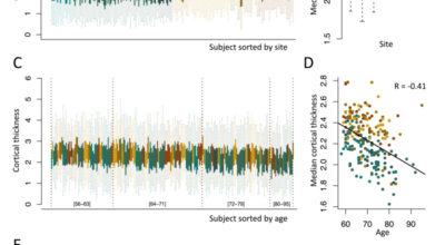
Harmonization of cortical thickness measurements across scanners
With the proliferation of multi-site neuroimaging studies, there is a greater need for handling non-biological variance introduced by differences in MRI scanners and acquisition protocols. Such unwanted sources of variation, […]
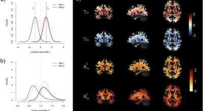
Harmonization of multi-site diffusion tensor imaging data
Diffusion tensor imaging (DTI) is a well-established magnetic resonance imaging (MRI) technique used for studying microstructural changes in the white matter. As with many other imaging modalities, DTI images suffer […]
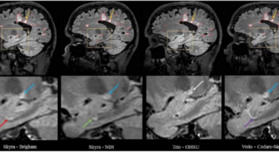
Volumetric Analysis from a Harmonized Multisite Brain MRI Study of a Single Subject with Multiple Sclerosis
MR imaging can be used to measure structural changes in the brains of individuals with multiple sclerosis and is essential for diagnosis, longitudinal monitoring, and therapy evaluation. The North American […]
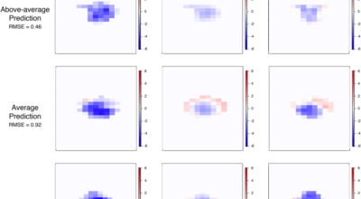
Predicting Degree and Spatial Pattern of MS Lesion Tissue Recovery
PREVAIL: In this work, our lab developed a model for predicting the degree and spatial pattern of MS lesion tissue recovery. The model is based solely on lesion presentation at […]